ACT Science : Chemistry
Study concepts, example questions & explanations for ACT Science


All ACT Science Resources
Example Questions
Example Question #51 : Chemistry

According to the graph at what temperature, in degrees celsius, are the solubilities for 

Between 30 and 40
Between 10 and 20
Between 60 and 70
Between 70 and 80
Between 40 and 50
Between 40 and 50
On the graph 

Example Question #52 : How To Find Data Representation In Chemistry
Based on the data in the graph, at 

Greater than

Greater than

Between 

Between 

Less than

Greater than
Assuming that the continuous positive slope of 


Example Question #52 : Chemistry
The table lists some of the properties of row 2 elements in the periodic table.

What conclusion can be drawn from the data in regards to atomic radius in row 2 elements on the periodic table?
An element with a low atomic radius will be a metal
An element with a high atomic radius will have high electronegativity
An element with a low atomic radius will have low electonegativity
An element with a high atomic radius will be a metal
An element with a low atomic radius will have a low atomic number
An element with a high atomic radius will be a metal
From studying the table it can be seen that the elements with the top two highest atomic radii are metals. As the atomic radius decreases elements are non-metals.
Example Question #54 : How To Find Data Representation In Chemistry
The table lists some of the properties of row 2 elements in the periodic table.
Which of the following graphs best represents the relationship between atomic radius and electronegativity for row 2 elements?


The table shows that as the atomic radius increases the electronegativity decreases. However, the atomic radius does not increase in continuous increments but in fact increases in larger subsequent increments as electronegativity decreases. Therefore, the graph will contain a curved line with a negative slope instead of a straight line.
Example Question #53 : Chemistry
Assuming that all of the weight lost or gained were solely from fat, determine the calories lost for the group subjected to only Inhibitor II. The energy density of fat is 9 calories per gram.
14,000 calories
37,000 calories
22,000 calories
27,000 calories
31,000 calories
27,000 calories
The approximate weight loss is about 3 kg for the Inhibitor II group. Therefore

Example Question #54 : Chemistry
Compute the mass of the fatty acid produced if inhibitor II were present in a sample of FAS and the experiment were to run for 20 seconds. Assume the fatty acid being produced is oleic acid, with a molecular weight of 282 grams.
.0056 grams
.0014 grams
.0093 grams
.0021 grams
.0087 grams
.0056 grams
Given that the rate of FAS with Inhibitor II present is 


Moles produced =

We can then compute the molecular weight by simply multiplying the number of moles by the molecular weight (282 grams), yielding 0.0056 grams.
Example Question #55 : Chemistry
A student performed the following procedures to study various photosynthetic pigments (light-absorbing chemicals) in tree leaves and the wavelengths of light they absorb.
Experiment 1:
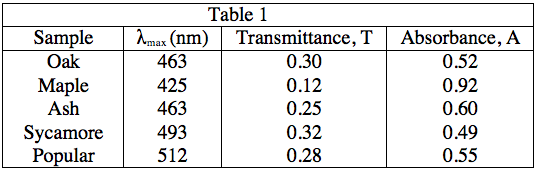
The student obtained samples of leaves from oaks, maples, ashes, sycamores, and poplars. Each leaf sample was ground separately with a mortar and pestle to release the pigments, and then each sample was suspended in water to make a colored solution of the pigment. The student then measured the absorption spectrum (a graph of how much light is absorbed by a pigment at varying wavelengths of light) of each solution in a device called a spectrophotometer. The setup of a spectrophotometer is shown below in Diagram 1.
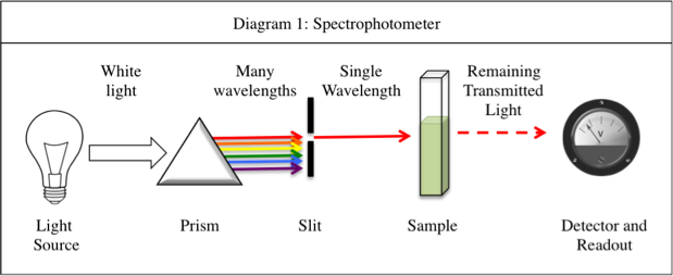
The light source emits white light, which is split into its various wavelengths by the prism. Next, a slit, which can be moved up or down to select a particular wavelength, is used to transmit just a single wavelength to the sample. The sample absorbs a fraction of this light that is characteristic to the pigment in the sample, and the rest is transmitted to the detector for a readout. Using the spectrophotometer, the student found the λmax (the wavelength of light in nanometers (nm) that the pigment absorbs most intensely, for each sample) and recorded the results in Table 1. Table 1 also shows the transmittance and absorbance values at λmax. Transmittance, T, is defined as the fraction of light, expressed as a decimal, which passes through the sample. Absorbance, A, is given by:
A = –log(T) or 10–A = T
Experiment 2:
A student is given a leaf from an unknown source. She crushes and extracts the pigment according to the procedure in Experiment 1. Measuring the absorbance spectrum in the spectrophotometer produces the following readout, shown in Diagram 2.
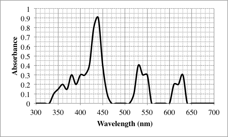
Diagram 2
Which of the following leaves most likely have the same pigment in high quantities?
Maple and Sycamore
Oak and Ash
Maple and Ash
Oak and Sycamore
Oak and Ash
The description of Experiment 1 states that λmax is a value characteristic of a particular pigment. Because λmax = 436nm for both Oak and Ash leaves, it can be assumed that this is because both leaves contain large amounts of the same pigment.
Example Question #56 : Chemistry
A student performed the following procedures to study various photosynthetic pigments (light-absorbing chemicals) in tree leaves and the wavelengths of light they absorb.
Experiment 1:
The student obtained samples of leaves from oaks, maples, ashes, sycamores, and poplars. Each leaf sample was ground separately with a mortar and pestle to release the pigments, and then each sample was suspended in water to make a colored solution of the pigment. The student then measured the absorption spectrum (a graph of how much light is absorbed by a pigment at varying wavelengths of light) of each solution in a device called a spectrophotometer. The setup of a spectrophotometer is shown below in Diagram 1.
The light source emits white light, which is split into its various wavelengths by the prism. Next, a slit, which can be moved up or down to select a particular wavelength, is used to transmit just a single wavelength to the sample. The sample absorbs a fraction of this light that is characteristic to the pigment in the sample, and the rest is transmitted to the detector for a readout. Using the spectrophotometer, the student found the λmax (the wavelength of light in nanometers (nm) that the pigment absorbs most intensely, for each sample) and recorded the results in Table 1. Table 1 also shows the transmittance and absorbance values at λmax. Transmittance, T, is defined as the fraction of light, expressed as a decimal, which passes through the sample. Absorbance, A, is given by:
A = –log(T) or 10–A = T
Experiment 2:
A student is given a leaf from an unknown source. She crushes and extracts the pigment according to the procedure in Experiment 1. Measuring the absorbance spectrum in the spectrophotometer produces the following readout, shown in Diagram 2.
Diagram 2
What is λmax, in nanometers, for the leaf in Experiment 2?
0.9
630
440
0.6
440
Experiment 1 states that λmax is the wavelength at which light is absorbed most intensely. Thus, we can look for the wavelength, found on the x-axis of Diagram 2, that produces the highest absorbance, found on the y-axis. This value is 440 nm, which produces an absorbance of 0.9.
Example Question #57 : How To Find Data Representation In Chemistry
A student wanted to study the kinetics, or rates of a chemical reaction based on the concentrations of its reactants and products, of the reaction shown below.
}+2KMnO_4_{(aq)}+3H_2SO_4_{(aq)}\rightarrow")
}+2MnSO_4_{(aq)}+K_2SO_4_{(aq)}+8H_2O_{(l)}")
This reaction is easy to monitor using a spectrophotometer, which measures how much light of a particular wavelength is absorbed by a solution. The deep purple potassium permanganate, or 

Experiment 1:
The student constructed a standard curve, or a graph of the absorbance of solutions of varying concentrations of potassium permanganate, to quantify the relationship between concentration and absorbance. To prepare five sample of increasing concentration, he labeled five test tubes A, B, C, D, and E, weighed out 0.1, 0.2, 0.3, 0.4, and 0.5 grams of potassium permanganate into each, respectively, and added 1 milliliter (mL) of water to each test tube to dissolve. Then, he used the spectrophotometer to determine the absorbance at 550 nm of each sample. The data is graphed in Figure 1 below.
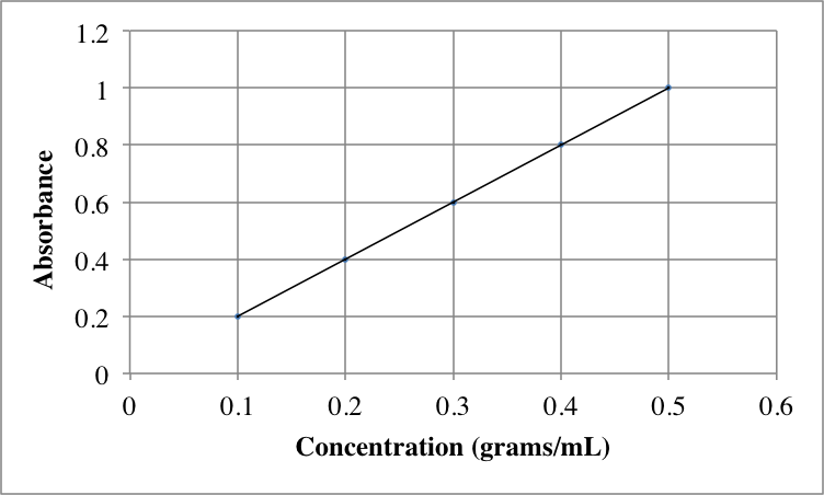
Figure 1
Experiment 2:
The student then studied potassium permanganate in the presence of oxalic acid, 
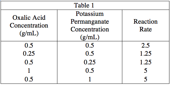
A sample solution of potassium permanganate in 1 milliliter of water was placed in a spectrophotometer and evaluated for its absorbance at 550 nm. It gave an absorbance of 0.3. How many grams of potassium permanganate were dissolved in the sample?


Figure 1 shows the relationship between absorbance and concentration of potassium permanganate. Look for the absorbance value of 0.3 on the vertical axis and see what concentration value on the horizontal axis would produce such an absorbance. The answer is 1.5 grams/mL. As the sample in this problem was dissolved in the same amount of water as in Experiment 1, we can assume that 0.15 grams were dissolved in this sample.
Example Question #56 : How To Find Data Representation In Chemistry
A student wanted to study the kinetics, or rates of a chemical reaction based on the concentrations of its reactants and products, of the reaction shown below.
This reaction is easy to monitor using a spectrophotometer, which measures how much light of a particular wavelength is absorbed by a solution. The deep purple potassium permanganate, or
Experiment 1:
The student constructed a standard curve, or a graph of the absorbance of solutions of varying concentrations of potassium permanganate, to quantify the relationship between concentration and absorbance. To prepare five sample of increasing concentration, he labeled five test tubes A, B, C, D, and E, weighed out 0.1, 0.2, 0.3, 0.4, and 0.5 grams of potassium permanganate into each, respectively, and added 1 milliliter (mL) of water to each test tube to dissolve. Then, he used the spectrophotometer to determine the absorbance at 550 nm of each sample. The data is graphed in Figure 1 below.
Figure 1
Experiment 2:
The student then studied potassium permanganate in the presence of oxalic acid, 
Which of the following graphs could potentially generated if absorbance at 550 nm was graphed over time for a mixture of potassium permanganate and oxalic acid?

Purple graph
Blue graph
Green graph
Red graph
Red graph
As the absorbance is monitored at 550 nm, which will observe the behavior of potassium permanganate, a reactant, we know the absorbance should decrease as time goes on. The concentration of a reactant will decrease as it is used up by the reaction. Thus, the answer must be the red graph, as it is the only graph that shows consistently decreasing absorbance.


All ACT Science Resources

