ACT Science : How to find experimental design in biology
Study concepts, example questions & explanations for ACT Science


All ACT Science Resources
Example Questions
Example Question #31 : How To Find Experimental Design In Biology
A scientific experiment is conducted to test if calcium can affect gene regulation. Scientists hypothesize that high levels of calcium would interact with the proteins Cs3 and Gfy, which in turn would increase the transcription of genes F4597 and BC392. The experiment procedure is summarized below.
- Isolate the genes F4597 and BC392.
- Create a vector within yeast cells containing the two genes
- Culture yeast cells
- Grow yeast cells in different growth mediums—one medium lacking calcium (plate A), and one medium with supplemented calcium (plate B)
What could be changed to strengthen the design of the experiment?
Use another substance instead of calcium as the independent variable
Looking at just one protein-gene interaction
Take out the step looking at protein interaction and focus just on the effects of calcium on the F4597 and BC392 gene activity
Nothing could be changed to strengthen the design experiment.
Having a control plate
Having a control plate
Having a control plate would allow the researchers to know the vectors were done correctly, and that the yeast cells were healthy and capable of growing regardless of calcium variabilty.
Example Question #543 : Biology
In the 1980’s, an epidemic of bovine spongiform encephalopathy, or mad cow disease, swept through cattle herds in the United Kingdom. Scientists and veterinarians were troubled and had a difficult time managing the disease because it spread from one animal to another, and behaved differently than other diseases in the past.
When infectious material from affected animals was treated with high levels of radiation, for example, the material remained infectious. All known bacteria or viruses that carry disease would have been killed by such a treatment. Additionally, some animals developed the disease without first being exposed to sick animals. Perhaps most frustratingly, among those animals that are exposed before becoming sick, it can take many years after exposure for illness to appear.
There quickly emerged two distinct explanations for the disease.
Scientist 1:
Mad cow disease is unlike any disease we have handled before. It is increasingly clear that the best explanation for the disease’s dynamics involve proteins, called the protein-only hypothesis. These protein molecules are likely causative of the disease, and they lack any DNA or RNA. It is damage to these DNA or RNA molecules that kills bacteria or viruses when exposed to high levels of radiation. The most important observations that made scientists consider a unique, protein-only model for this disease involved its resistance to radiation. Remarkably, this would be the first example of an infectious agent copying itself without DNA or RNA to mediate the process.
Moreover, some animals develop the disease spontaneously, without physically being infected by another animal. This suggests that internal disorder among protein molecules is a potential route to developing disease, and may be accelerated by exposure to other sick animals.
In fact, this is consistent with the proposed mechanism. It is likely that proteins fold incorrectly, and then influence proteins around them to take on this errant conformation. Some proteins may fold incorrectly by chance, which explains spontaneous disease development. It also explains the long course of disease, as it takes many years for enough proteins to fold incorrectly and result in observable disease.
Scientist 2:
The suggestion that mad cow disease is caused exclusively by protein, in the absence of DNA or RNA, is such a dramatic departure from accepted biological processes that it warrants careful scrutiny. Additionally, other more conventional explanations should be thoroughly investigated before coming to such a conclusion.
Some scientists have shown that very small particles resembling viruses are visible in infectious material under powerful microscopes. Additionally, these viruses are consistent in size and shape with known, highly radiation-resistant viruses called polyomaviruses. It takes much higher-than-typical doses of radiation to cause enough DNA damage to inactivate these viruses.
The observation that mad cow disease occurs spontaneously in some animals is also explained by the viral explanation. Many viruses exist in animals and humans for years, undetected and no causing any observable disease. Sickness or stress can make these viruses reactivate, offering the illusion of spontaneous illness. All of these observations are consistent with the viral hypothesis.
Suppose a scientist wishes to conduct an experiment to ascertain the presence of DNA or RNA in a sample from a cow with mad cow disease. He decides to compare his sample to a sample that is known to contain DNA and subject both to the same test. This known sample is best described as:
An experimental group
A balancing group
A negative control group
An outlier group
A positive control group
A positive control group
Control groups provide a comparison to an experimental group. In this case, the goal is to compare the group that is of interest, the experimental sample from the cow, to a known standard, the DNA positive control. Consider if we left this positive control off of our experiment, and our detection equipment was broken. We would get a false negative result. By including a known positive control, we would know that this negative result was erroneous.
Example Question #545 : Biology
Clostridium botulinum is a bacterial organism that can cause disease in people after eating improperly canned foods. As a result of this risk, canning foods involves bringing contents to high pressures and temperatures, thus killing the inactive form of Clostridium, called a spore.
Table 1 shows the ability of a scientist to detect spores as a function of the peak temperature and pressure reached during the process used for canning green beans.
|
Peak Temperature |
Peak Pressure |
Spores/Cubic Millimeter |
|
100 C |
50 PSI |
5 |
|
100 C |
100 PSI |
3 |
|
150 C |
50 PSI |
2 |
|
150 C |
100 PSI |
1 |
Table 2 shows the infectious dose of spores per cubic millimeter necessary to cause illness in four populations.
|
Population |
Minimum Concentration of Spores |
|
Children <1 Year |
1 |
|
Children 1-4 Years |
1 |
|
Children 5-10 years |
4 |
|
Children > 10 years and Adults |
8 |
A scientist discovers that, despite adhering to appropriate canning methods as described above, an outbreak of disease due to Clostridium botulinum has taken place in a Minnesota school. She visits the school and collects food samples to determine the cause of the outbreak. While compiling data at the school, she discovers that there are an increasing number of cases of a new strain of Clostridium botulinum. Upon investigation, the scientist finds that all children who attend the school are older than 5 years of age.
During her investigation of the outbreak, the scientist compared the symptoms of patients with the new strain of Clostridium, to patients with the original strain. She noticed signicificantly greater rates of diarrhea with the new strain. In this example, the group of patients with the original strain is serving as a:
Experimental group
Control group
Confounder group
Variable group
Modifier group
Control group
An experimental control group is a group that differs from an experimental group in just one way, the question being studied. Here, the original group and the control group are ideally identical in all respects, except in the strain of Clostridium botulinum with which they are infected.
Example Question #32 : How To Find Experimental Design In Biology
Understanding the biological features of different bacteria that allow them to grow in unwelcoming environments is necessary to treat and prevent human disease. Modern scientific laboratories, such as those in major hospitals, take blood, urine, and mucus samples from patients and culture them for bacterial growth. During the culturing process, laboratory technicians stain the growing bacteria for a component of their cell wall, the structure that provides shape and rigidity to the bacterium, through a process called Gram staining. Bacteria are typically classified as Gram Positive or Gram Negative, a distinction that is important in selecting the most effective antibiotic for treatment. Gram Positive bacteria appear purple under a microscope, while Gram Negative bacteria appear red. However, some bacteria do not Gram Stain and cannot be seen under a microscope when prepared this way.
Technicians also grow the bacteria on various types of plates containing special growth nutrients to determine which bacteria are causing a specific illness. If a bacterium is able to grow on a selective plate, meaning a plate that contains additional nutrients required for a specific bacterium to grow if it is present in the culture, doctors are able to determine the exact cause of a patient’s illness and prescribe targeted antibiotics to eliminate the infection. Bacteria that commonly cause human illness, their growth requirements, and their appearance on specific growth media are presented below in Table 1.
Table 1

Scientists can take the bacteria cultured on the plate and further analyze their enzymes. Three enzymes—urease, catalase, and beta-lactamase—are important for bacterial survival against the human immune system. Urease is responsible for producing urea, a basic molecule that can counteract the bactericidal (bacteria-killing) activity of stomach acid. Catalase, on the other hand, helps bacteria neutralize toxic substances released from human immune cells, allowing them to survive oxidative stress in high-oxygen areas. Finally, beta-lactamase allows Gram Positive bacteria to break down antibiotics called penicillins. While this ability to break down penicillin and its related antibiotic ampicillin was not initially present, bacteria, especially E. coli, have adapted by developing the new enzyme beta-lactamase that opens the ring responsible for penicillin’s bactericidal activity, rending the antibiotic ineffective. This and other examples of antibiotic resistance are becoming more common and are making treatment of serious human diseases very challenging.
What is the most likely reason that some organisms will not Gram stain?
Catalase is present.
They do not have a cell wall.
Beta-Lactamase is present.
Urease is present.
They do not have a cell wall.
This question asks us specifically about the gram staining procedure that is described in the first paragraph. The procedure described says the stain marks a cell wall. Thus, if a cell will not gram stain, it most likely does not contain a cell wall. The other answer choices do not impact the gram staining procedure described in the passage.
Example Question #33 : How To Find Experimental Design In Biology
Understanding the biological features of different bacteria that allow them to grow in unwelcoming environments is necessary to treat and prevent human disease. Modern scientific laboratories, such as those in major hospitals, take blood, urine, and mucus samples from patients and culture them for bacterial growth. During the culturing process, laboratory technicians stain the growing bacteria for a component of their cell wall, the structure that provides shape and rigidity to the bacterium, through a process called Gram staining. Bacteria are typically classified as Gram Positive or Gram Negative, a distinction that is important in selecting the most effective antibiotic for treatment. Gram Positive bacteria appear purple under a microscope, while Gram Negative bacteria appear red. However, some bacteria do not Gram Stain and cannot be seen under a microscope when prepared this way.
Technicians also grow the bacteria on various types of plates containing special growth nutrients to determine which bacteria are causing a specific illness. If a bacterium is able to grow on a selective plate, meaning a plate that contains additional nutrients required for a specific bacterium to grow if it is present in the culture, doctors are able to determine the exact cause of a patient’s illness and prescribe targeted antibiotics to eliminate the infection. Bacteria that commonly cause human illness, their growth requirements, and their appearance on specific growth media are presented below in Table 1.
Table 1
Scientists can take the bacteria cultured on the plate and further analyze their enzymes. Three enzymes—urease, catalase, and beta-lactamase—are important for bacterial survival against the human immune system. Urease is responsible for producing urea, a basic molecule that can counteract the bactericidal (bacteria-killing) activity of stomach acid. Catalase, on the other hand, helps bacteria neutralize toxic substances released from human immune cells, allowing them to survive oxidative stress in high-oxygen areas. Finally, beta-lactamase allows Gram Positive bacteria to break down antibiotics called penicillins. While this ability to break down penicillin and its related antibiotic ampicillin was not initially present, bacteria, especially E. coli, have adapted by developing the new enzyme beta-lactamase that opens the ring responsible for penicillin’s bactericidal activity, rending the antibiotic ineffective. This and other examples of antibiotic resistance are becoming more common and are making treatment of serious human diseases very challenging.
A technician stains a slide using the gram stain procedure and sees nothing upon looking under the microscope. Which growth medium could be required to determine is a particular bacterium is causing disease?
Lactose
Sheep's blood
None of the other answers
Chocolate agar
Sheep's blood
This question describes a technician that has completed a gram stain, yet sees nothing under the microscope. Using the provided table, we can see that one bacterium in particular, H. pylori, does not gram stain. We could use sheep's blood to determine if H. pylori is present.
Example Question #34 : How To Find Experimental Design In Biology
Understanding the biological features of different bacteria that allow them to grow in unwelcoming environments is necessary to treat and prevent human disease. Modern scientific laboratories, such as those in major hospitals, take blood, urine, and mucus samples from patients and culture them for bacterial growth. During the culturing process, laboratory technicians stain the growing bacteria for a component of their cell wall, the structure that provides shape and rigidity to the bacterium, through a process called Gram staining. Bacteria are typically classified as Gram Positive or Gram Negative, a distinction that is important in selecting the most effective antibiotic for treatment. Gram Positive bacteria appear purple under a microscope, while Gram Negative bacteria appear red. However, some bacteria do not Gram Stain and cannot be seen under a microscope when prepared this way.
Technicians also grow the bacteria on various types of plates containing special growth nutrients to determine which bacteria are causing a specific illness. If a bacterium is able to grow on a selective plate, meaning a plate that contains additional nutrients required for a specific bacterium to grow if it is present in the culture, doctors are able to determine the exact cause of a patient’s illness and prescribe targeted antibiotics to eliminate the infection. Bacteria that commonly cause human illness, their growth requirements, and their appearance on specific growth media are presented below in Table 1.
Table 1
Scientists can take the bacteria cultured on the plate and further analyze their enzymes. Three enzymes—urease, catalase, and beta-lactamase—are important for bacterial survival against the human immune system. Urease is responsible for producing urea, a basic molecule that can counteract the bactericidal (bacteria-killing) activity of stomach acid. Catalase, on the other hand, helps bacteria neutralize toxic substances released from human immune cells, allowing them to survive oxidative stress in high-oxygen areas. Finally, beta-lactamase allows Gram Positive bacteria to break down antibiotics called penicillins. While this ability to break down penicillin and its related antibiotic ampicillin was not initially present, bacteria, especially E. coli, have adapted by developing the new enzyme beta-lactamase that opens the ring responsible for penicillin’s bactericidal activity, rending the antibiotic ineffective. This and other examples of antibiotic resistance are becoming more common and are making treatment of serious human diseases very challenging.
Which of the following bacteria is most likely to produce urease?
S. pneumoniae
H. pylori
H. influenzae
E. coli
H. pylori
Using the last paragraph as a guide, we can see that urease is able to combat the acidic environment of the stomach. Thus, we could predict that H. pylori, which causes stomach infections, would most likely produce urease to combat the acidic environment of the stomach.
Example Question #35 : How To Find Experimental Design In Biology
Understanding the biological features of different bacteria that allow them to grow in unwelcoming environments is necessary to treat and prevent human disease. Modern scientific laboratories, such as those in major hospitals, take blood, urine, and mucus samples from patients and culture them for bacterial growth. During the culturing process, laboratory technicians stain the growing bacteria for a component of their cell wall, the structure that provides shape and rigidity to the bacterium, through a process called Gram staining. Bacteria are typically classified as Gram Positive or Gram Negative, a distinction that is important in selecting the most effective antibiotic for treatment. Gram Positive bacteria appear purple under a microscope, while Gram Negative bacteria appear red. However, some bacteria do not Gram Stain and cannot be seen under a microscope when prepared this way.
Technicians also grow the bacteria on various types of plates containing special growth nutrients to determine which bacteria are causing a specific illness. If a bacterium is able to grow on a selective plate, meaning a plate that contains additional nutrients required for a specific bacterium to grow if it is present in the culture, doctors are able to determine the exact cause of a patient’s illness and prescribe targeted antibiotics to eliminate the infection. Bacteria that commonly cause human illness, their growth requirements, and their appearance on specific growth media are presented below in Table 1.
Table 1
Scientists can take the bacteria cultured on the plate and further analyze their enzymes. Three enzymes—urease, catalase, and beta-lactamase—are important for bacterial survival against the human immune system. Urease is responsible for producing urea, a basic molecule that can counteract the bactericidal (bacteria-killing) activity of stomach acid. Catalase, on the other hand, helps bacteria neutralize toxic substances released from human immune cells, allowing them to survive oxidative stress in high-oxygen areas. Finally, beta-lactamase allows Gram Positive bacteria to break down antibiotics called penicillins. While this ability to break down penicillin and its related antibiotic ampicillin was not initially present, bacteria, especially E. coli, have adapted by developing the new enzyme beta-lactamase that opens the ring responsible for penicillin’s bactericidal activity, rending the antibiotic ineffective. This and other examples of antibiotic resistance are becoming more common and are making treatment of serious human diseases very challenging.
Assume a new growth medium was created that contained a mixture of sheep’s blood, lactose, and Factors X and V (chocolate agar) but was also supplemented with penicillin. What type of bacteria could likely be cultured on this new medium?
H. influenzae
E. coli
S. pneumoniae
B. cerrius
E. coli
The question asks us to identify the bacteria that would most likely grow on the selective medium that includes penicillin. The information we need to answer this question is contained in the last paragraph, where we see that E. coli is the only bacteria that is resistant to penicillin via the production of beta-lactamase.
Example Question #550 : Biology
A researcher studies the olfactory (scent-related) senses of the giant forest ant, Camponotus gigas. The researcher places 120 ants in a three-chambered cell. The cell has an end section with a cotton ball soaked in a saline solution and another end with a cotton ball soaked in a glucose solution. The ants are placed in the middle and timed for 15 minutes. Their initial and final positions in the cell are recorded (see Table 1). The researcher's null hypothesis states that the distribution of ants across the three chambers will be equal to one another. In other words, the glucose solution will have no effect and there will be no significant difference in the distribution of the insects.
Table 1
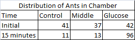
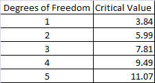
The researcher decides to perform a statistical test known as a Chi-squared test of independence to interpret the experiment's results. The test is performed by calculating a Chi-squared statistic by utilizing observed and expected values for distribution (see Table 2). If the sum of the Chi-squared test statistic is higher than the critical value, then the null hypothesis can be rejected. This indicates that the distribution of insects is not random and the variable in question has a pronounced effect on the subjects. A critical value is calculated by determining the degrees of freedom, which in this experiment is equal to the number of categories in the study minus one, and then locating the proper number on a table (see Table 3). There are three possible categories in this experiment: the glucose end, the control end (with the saline-solution soaked cotton ball), and middle portion of the chamber.
Table 2
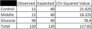
Table 3
If the null hypothesis is rejected for this study, which of the following would be most likely be true?
None of the other choices are correct.
The ants would demonstrate a pattern of distribution in the cell.
It is impossible to predict how the ants would be distributed.
The ants would be equally distributed throuhgout the cell's chambers.
The ants would demonstrate a pattern of distribution in the cell.
The null hypothesis is a stament of no difference, which means that if it were supported, then the ants should be equally distributed across the cell. If the null hypothesis were to be rejected, the ants would demonstrate a pattern of distribution in the cell.
Example Question #551 : Act Science
A researcher studies the olfactory (scent-related) senses of the giant forest ant, Camponotus gigas. The researcher places 120 ants in a three-chambered cell. The cell has an end section with a cotton ball soaked in a saline solution and another end with a cotton ball soaked in a glucose solution. The ants are placed in the middle and timed for 15 minutes. Their initial and final positions in the cell are recorded (see Table 1). The researcher's null hypothesis states that the distribution of ants across the three chambers will be equal to one another. In other words, the glucose solution will have no effect and there will be no significant difference in the distribution of the insects.
Table 1
The researcher decides to perform a statistical test known as a Chi-squared test of independence to interpret the experiment's results. The test is performed by calculating a Chi-squared statistic by utilizing observed and expected values for distribution (see Table 2). If the sum of the Chi-squared test statistic is higher than the critical value, then the null hypothesis can be rejected. This indicates that the distribution of insects is not random and the variable in question has a pronounced effect on the subjects. A critical value is calculated by determining the degrees of freedom, which in this experiment is equal to the number of categories in the study minus one, and then locating the proper number on a table (see Table 3). There are three possible categories in this experiment: the glucose end, the control end (with the saline-solution soaked cotton ball), and middle portion of the chamber.
Table 2
Table 3
In the Chi-squared test, what was the expected number of ants in each chamber?


Table 2 contains the Chi-squared information for this experiment. The column titled "Expected" shows that the researcher would expect a uniform distribution of the 120 ants across each of the cell's chambers. This indicates that there the expected number of ants in each cell is 40.
Example Question #32 : How To Find Experimental Design In Biology
A researcher studies the olfactory (scent-related) senses of the giant forest ant, Camponotus gigas. The researcher places 120 ants in a three-chambered cell. The cell has an end section with a cotton ball soaked in a saline solution and another end with a cotton ball soaked in a glucose solution. The ants are placed in the middle and timed for 15 minutes. Their initial and final positions in the cell are recorded (see Table 1). The researcher's null hypothesis states that the distribution of ants across the three chambers will be equal to one another. In other words, the glucose solution will have no effect and there will be no significant difference in the distribution of the insects.
Table 1
The researcher decides to perform a statistical test known as a Chi-squared test of independence to interpret the experiment's results. The test is performed by calculating a Chi-squared statistic by utilizing observed and expected values for distribution (see Table 2). If the sum of the Chi-squared test statistic is higher than the critical value, then the null hypothesis can be rejected. This indicates that the distribution of insects is not random and the variable in question has a pronounced effect on the subjects. A critical value is calculated by determining the degrees of freedom, which in this experiment is equal to the number of categories in the study minus one, and then locating the proper number on a table (see Table 3). There are three possible categories in this experiment: the glucose end, the control end (with the saline-solution soaked cotton ball), and middle portion of the chamber.
Table 2
Table 3
The researcher designs a follow-up experiment that uses five categories. How many degrees of freedom will this experiment possess?


The passage states that the degrees of freedom are calculated by taking the number of catagories in an experiment and subtracting one. The researcher plans to use five categories in the new experiement.

Therefore, there will be four degrees of freedom in this new experiment.

Certified Tutor

Certified Tutor

All ACT Science Resources

